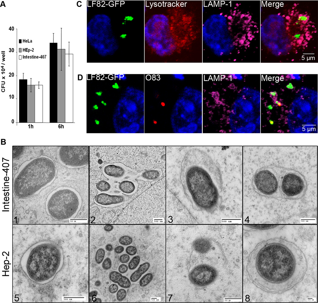
Figure 3. AIEC LF82 bacteria interact with autophagy in epithelial cell lines.
A. Intracellular replication of AIEC LF82 bacteria at 1 h and 6 h post-infection within HeLa, Hep-2 and Intestine-407 epithelial cells. Results are expressed as mean numbers +/− SEM of colony forming units (CFU) per well. Each point is the mean of at least three separate experiments. B. Ultrastructural analysis of Intestine-407 and Hep-2 epithelial cells infected with AIEC strain LF82 for 6 h by transmission electron microscopy showing various intracellular compartments for intracellular LF82 bacteria: single bacteria in monolayer membrane vacuoles (1 and 5), several bacteria in a single membrane vacuole (2 and 6), bacteria in damaged vacuole (3 and 7) and bacteria in a multilamellar membrane vacuole also containing sequestered cytoplasm (4 and 8). C. Confocal microscopic examinations of AIEC LF82 GFP-infected Hela cells at 6 h post-infection labelled with lysotracker (red) and for LAMP-1 (purple) to visualize the vacuoles. D. Confocal analysis after permeabilization of plasma membrane with digitonin and labelling of free cytosolic AIEC LF82 with O83 antibodies (red) and of vacuoles with LAMP-1 antibodies (purple).

