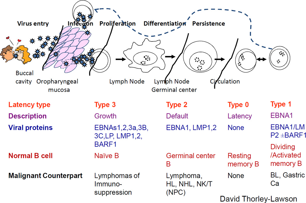
Figure 1. Epstein-Barr virus life cycle, latency states and lymphoma.
The Epstein-Barr virus (EBV) life cycle involves at least five distinct stages and four are associated with disease. During primary infection, EBV infects naïve B-cells and expresses its entire latency gene complex, including 10 proteins and 2 small RNAs (type III latency). Type III latency drives B-cell transformation and proliferation, but because the cells are highly immunogenic, they are rapidly eliminated by EBV-specific T-cells. The virus survives in B-cells by downregulating its immunogenic proteins in two phases. Initially B-cells enter lymphoid follicles where they proliferate and express only three viral proteins (type III latency). Finally they exit the lymph node and downregulate viral proteins altogether (type 0 latency), and thus are invisible to the immune response. If circulating infected B-cells divide homeostatically they express a single viral protein (EBNA1, type I latency) that ensures that the virus genome divides with the cell genome. When infected B-cells circulate through the oropharynx they transfer the virus to epithelial cells, where it is replicated to infect new hosts by salivary transfer and where it infects new B cells to maintain the infected B-cell pool. With the exception of type I latency, each latency state is observed in specific types of lymphoma. Abbreviations: EBNA, Epstein-Barr nuclear antigen; EBV, Epstein-Barr virus

