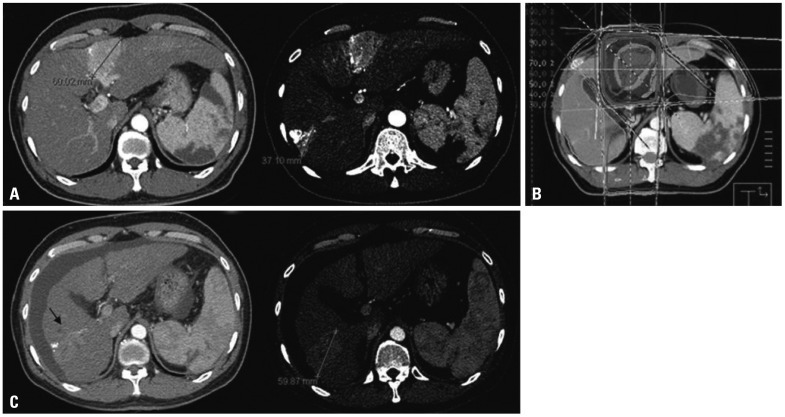
Fig. 1.
Illustrations of a patient in the primary group who achieved in-field CR but had out-field progression in the liver. (A) The pretreatment computed tomography (CT) scan shows two lesions. The lesion located in the segment 6 was treated with transarterial chemoembolization. (B) Axial dose distribution of 3-D conformal radiotherapy (RT). The lesion in the segment 4 was treated with RT, and (C) 1 month after completion of RT, a follow-up CT scan shows disappearance of the lesion in the segment 4. Unfortunately, the lesion in the segment 6 had progressed. CR, complete remission.

