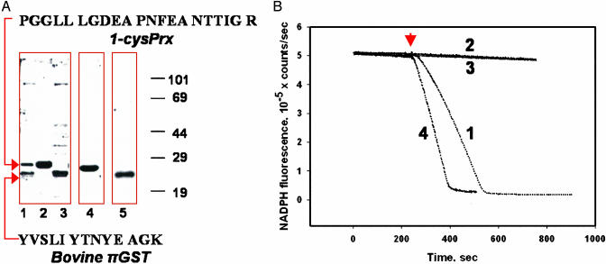
Fig. 1.
Activation of 1-cysPrx by incubation with πGST. (A) Lanes 1-3, SDS/PAGE under reducing conditions (2 mM DTT) and stained with Simply Blue. Lane 1, starting material for GST Trap column; lane 2, concentrated eluate from the column; lane 3, concentrated retentate after elution of the column with 10 mM GSH. Arrows indicate the bands that were analyzed by MS. The sequences of the analyzed peptides and their identification by blast analysis are shown. Western blot: lane 4, lane 2 probed with 1-cysPrx mAb; lane 5, lane 3 probed with πGST pAb. (B) Peroxidase activity by NADPH/GSH reductase/GSH-coupled assay with PLPCOOH (addition indicated by arrow) as substrate. Activities were 5.0 ± 0.4 μmol/min/mg of protein for partially purified bovine lung enzyme (trace 1), zero for purified 1-cysPrx after storage for 1 week at 4°C (trace 2) and for πGST alone (trace 3), and 4.5 ± 0.4 μmol/min/mg of protein (mean ± SE, n = 3) after GSH-saturated πGST addition to the purified inactive protein (1:1 molar ratio) (trace 4).

