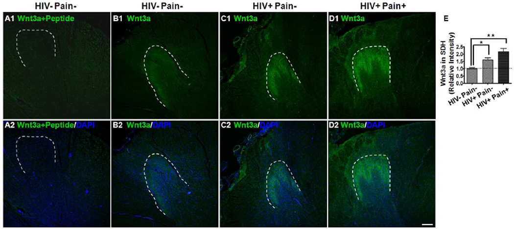
Fig. 2.
Spatial distribution of Wnt3a in the SDH. (A1–A2) Immunostaining results (HIV− pain− patients) with the antibody pre-blocked with Wnt3a peptide. (B–D) Immunostaining results of Wnt3a in the SDH of an HIV-negative subject (B1–B2), a ‘pain-negative’ HIV patient (C1–C2), and a ‘pain-positive’ HIV patient (D1–D2). Wnt3a signals formed a predominant band in the SDH, and the signals were hard to detect outside of the dorsal horn. (E) Intensity of Wnt staining in the SDH. The Wnt signal was markedly increased in the SDH of ‘pain-positive’ HIV patients, compared with ‘pain-negative’ HIV patients or HIV-negative subjects (mean ± SEM; * p<0.05; ** p<0.01; one-way ANOVA). DAPI (Blue) staining was included to visualize all cells. Dashed lines were drawn to indicate the SDH regions. Scale bar: 400 µm.

