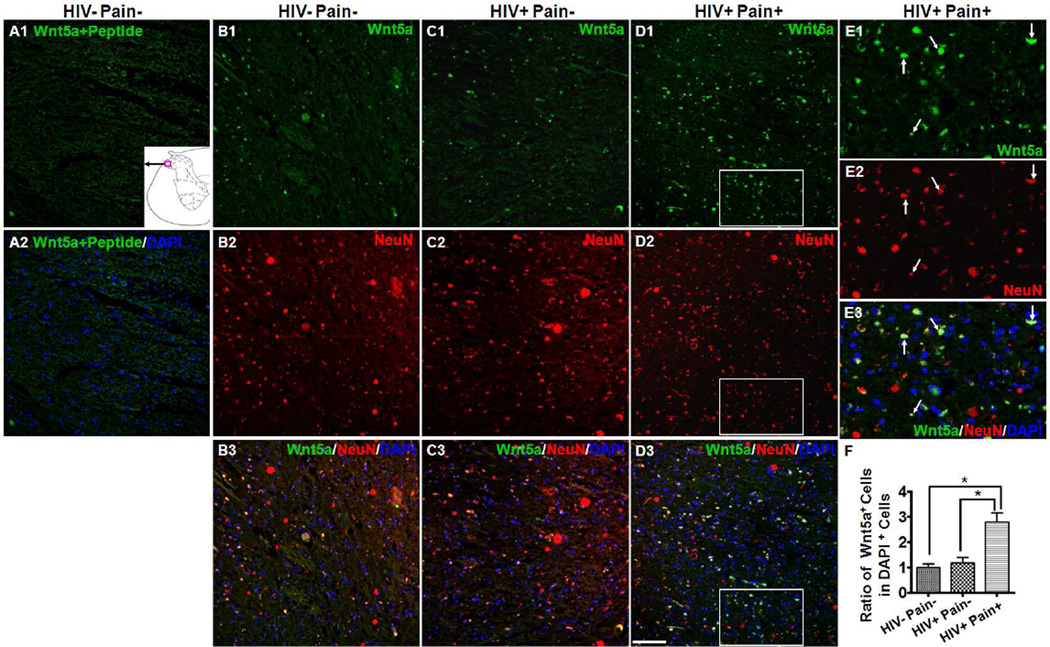
Fig. 4.
Cellular localization of Wnt5a in the SDH. (A1–A2) Immunostaining results with Wnt5a antibody pre-blocked with Wnt5a peptide (HIV− pain− subjects). (B–D) Double staining of Wnt5a (Green) and NeuN (Red) in the SDH of an HIV-negative subject (B1–B3), ‘pain-negative’ HIV patients (C1–C3), and ‘pain-positive’ HIV patients (D1–D3). Wnt5a signals formed bright spots in the SDH and were found in NeuN-labeled cell bodies. (E1–E3) Higher power images of the box regions in (D1–D3). Most Wnt5a signals were co-localized with NeuN in the neuronal cell bodies (arrows). (F) Quantitative Wnt5a-postive cells in the SDH. Wnt5a-positive cells were significantly up-regulated in the SDH of ‘pain-positive’ HIV patients (mean ± SEM; * p<0.05; one-way ANOVA). Scale bar: 100 µm.

