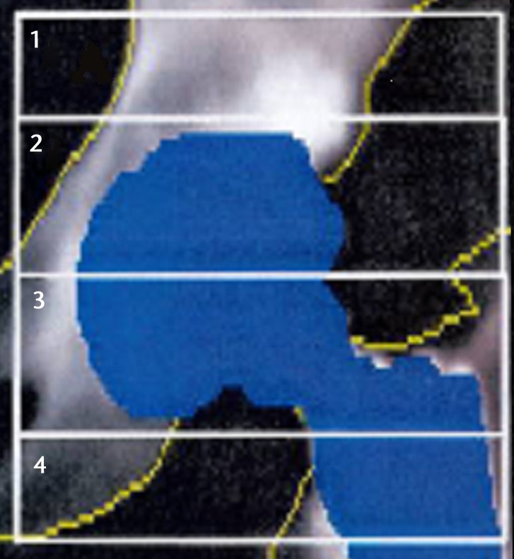
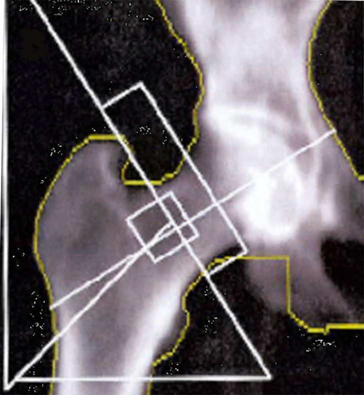
Figs. 2a - 2b.


Images showing the regions of interest for the assessment of bone mineral density in a) the affected hip and b) the contralateral hip.


Images showing the regions of interest for the assessment of bone mineral density in a) the affected hip and b) the contralateral hip.