Fig. 1.
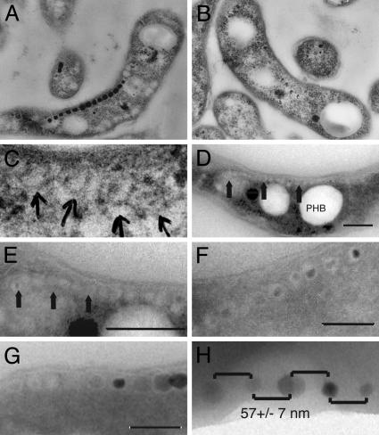
Characterization of magnetosome dynamics by electron microscopy. (A) TEM image of an ultrathin section of Magnetospirillum sp. AMB-1 grown under iron-rich conditions and embedded in epoxy resin. A chain of magnetite crystals can be observed in the cells. (B) AMB-1 cells grown without iron do not contain magnetite chains. (C) Higher magnification of the cell shown in B reveals potential empty magnetosome vesicles. (D) TEM image of cryo-ultrathin section of iron-starved AMB-1 showing a long chain of empty magnetosome vesicles. The three large white inclusions are polyhydroxybutyrate (PHB) granules. A magnetosome chain (arrows) is seen on top of these PHB granules. (E) Higher magnification of magnetosomes in D. Arrows point to magnetosome chains devoid of magnetite. (F) TEM image of a cryo-ultrathin section of AMB-1 cells 7 h after the addition of iron to iron-starved cultures. Small magnetite crystals show the early stages of crystal growth within vesicles having relatively uniform size and shape. It is evident that simultaneous nucleation and growth of magnetite crystals occurs within the full-sized vesicles. (G) TEM image of a cryo-ultrathin section of AMB-1 after 20 h of growth in iron-rich medium. Structures resembling empty magnetosomes are seen at the ends of chains of fully grown magnetite crystals. (H) Regular spacing of magnetite crystals 2 h after the addition of iron. Whole cells of AMB-1 were imaged in TEM without sectioning. The spacing between magnetite crystals is very even, with an average of 57 ± 7 nm. (Scale bars: 200 nm in D-G.)

