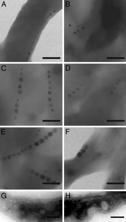
Fig. 3.
ΔmamA cells have a defect in activating their magnetosome vesicles. (A-F) TEM images of whole cells of WT and ΔmamA after the addition of iron to iron-starved cells. At 2 h both WT (A) and ΔmamA (B) cells have started synthesizing magnetite. The crystals get progressively larger at 3.5 h (C and D) and are full-sized after 21 h (E and F). At all times, however, WT cells have more crystals per chain than ΔmamA cells. (G and H) TEM images of cryo-ultrathin sections of ΔmamA cells grown in the absence (G) or presence (H) of iron, revealing the presence of chains of empty vesicles similar to WT (Fig. 1 E and F). (Scale bars: 200 nm.)

