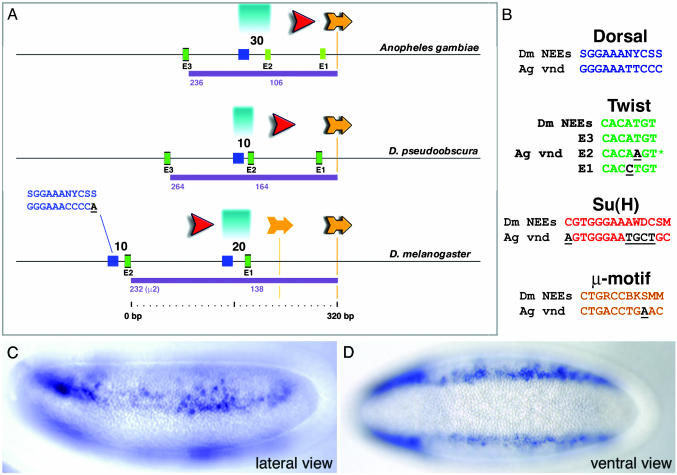
Fig. 4.
Anopheles vnd enhancer retains conserved NEE structure and function. (A) The mosquito vnd enhancer displays many of the organizational features seen for all of the Drosophila neurogenic enhancers. Comparison with the Drosophila pseudoobscura NEE sequence reveals an interesting intermediate compared to the former two. Both the Twist/Dorsal-like and Twist/μ distances are indicated for each sequence. For discussion of salient aspects of this organization, see text. (B) Alignments of Anopheles vnd sites to Drosophila NEE consensus sequences are depicted with mismatches underlined. The E2 sequence (asterisk) shown here overlaps a CACTTGT E-box in the opposite orientation. Lateral (C) and ventral (D) views of transgenic Drosophila embryos carrying the Anopheles vnd enhancer attached to the lacZ reporter gene are shown. Staining is detected in ventral regions of the neurogenic ectoderm.

