Fig. 1.
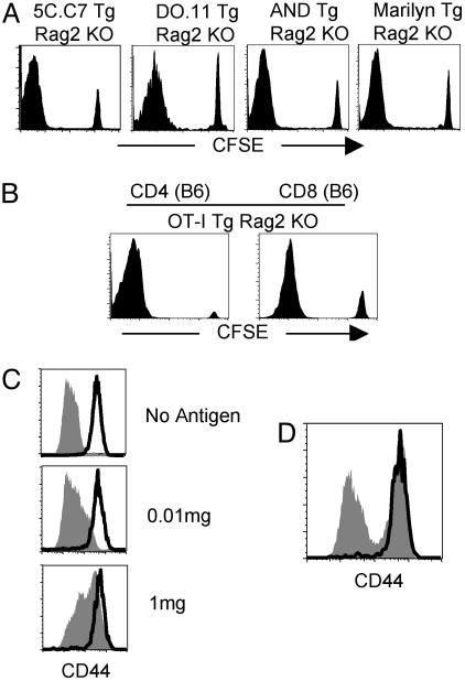
CD4 T cells of single specificity fail to block the proliferation of polyclonal naïve CD4 T cells. (A) A total of 2 × 10⁶ CFSE-labeled naïve CD4 T cells from Ly5.1 B10.A, BALB/c, and C57BL/10 mice were transferred into 5C.C7 Tg Rag2-/-, DO.11 Tg Rag2-/-, and AND Tg Rag2-/- or Marilyn Tg Rag2-/- mice, respectively. Shown is CFSE profile of transferred cells (gated on Ly5.1+, KJ1.26+, Vβ3-, and Vβ6- CD4+, respectively) at 7 days after transfer. (B) CFSE-labeled naïve Ly5.1 CD4 and CD8 T cells were transferred into OT-I Tg Rag1-/- mice. CFSE profile was measured at 7 days after transfer (gated on Ly5.1+ CD4+). (C) 5C.C7 Tg Rag2-/- mice received 2 × 10⁶ CFSE-labeled Ly5.1 CD4 T cells and were simultaneously implanted with a miniosmotic pump containing 0, 0.01, or 1 mg of cytochrome C protein. Lymph node CD4 T cells were analyzed for CD44 expression after 7 days of transfer. (D) Ly5.1 naïve CD4 T cells were transferred into 5C.C7 Tg Rag2-/- mice that had been immunized 60 days earlier by implantation of a miniosmotic pump containing 1 mg of cytochrome C protein. CD44 expression was measured 7 days after transfer. The bold line represents Ly5.1 cells and the filled area represents endogenous Tg cells. Experiments were repeated more than twice with similar results.

