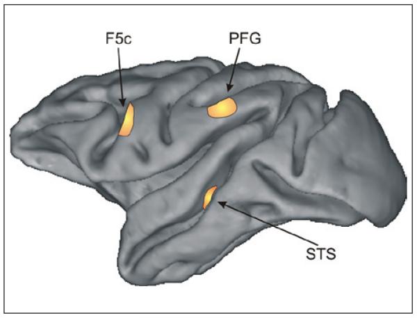
Figure 1.
Areas in the monkey brain involved in action perception. The figure shows a lateral view of the left hemisphere of a macaque brain. The three highlighted areas contain neurons that discharge during observation of bodily movements. More specifically, neurons in the superior temporal sulcus (STS) exhibit purely visual responses and do not respond during the execution of movements. Neurons responding both during the observation and execution of goal-directed movements (i.e., mirror neurons) were found in areas F5c and PFG.

