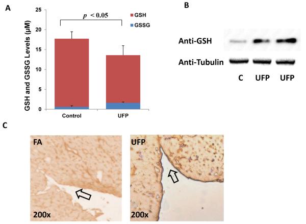
Fig. 2. UFP increased protein S-glutathionylation in HAEC.
(A) HAEC were treated with or without 50μg/mL of UFP for 6 hours, cellular levels of GSH and GSSG were measured as described in Methods. UFP decreased GSH level, but increased GSSG level. (n=4) (B) HAEC were treated with or without 50μg/mL of UFP for 6 hours and protein lysates were collected. Western blots with antibody against GSH revealed an increase in a dominant band to S-Glutathionylated Actin. The western blot with anti-Tubulin was performed as loading reference. (C) Sections of endocardium from LDLR-null mice exposed to filtered air (FA) or UFP for 10 weeks were stained with anti-GSH antibody for visualization of protein S-glutathionylation. UFP exposure led to a prominent staining in the endothelium of endocardium (Arrow).

