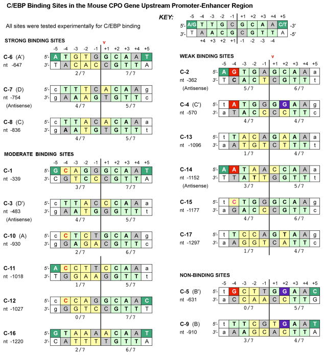
Figure 4. Comparative sequence analysis of C/EBP sites in the murine CPO upstream region.
Each double-stranded DNA site for C/EBP binding consists of two 5 bp half-sites centered on a dyad axis of symmetry (vertical double-lines). Boxes are shaded as follows: Light green: this nucleotide (NT) is identical to the NT shown to contact specific amino acids of C/EBP in the crystallographic studies of Miller et al (33). Yellow: Deviations from the NT shown in Miller et al, 2003. Dark green: A pyrimidine is the NT that occurs most commonly at position (+5) in natural promoters (31, 32). Red: purine at position −4 is sterically unfavorabe. Purple: a guanine is normally never observed at position +2, except in weak or nonbinding sites. Gray, the NT at this location is not critical for binding (33), and therefore not considered in the scoring system. Arrow, this nucleotide (+1) defines the location of the site in the mCPO promoter. Numerals beneath each 5 bp half-site: This is the consensus score, i.e., the number of NTs that match the theoretically perfect consensus sequence; 7 out of a possible 7 is perfect.

