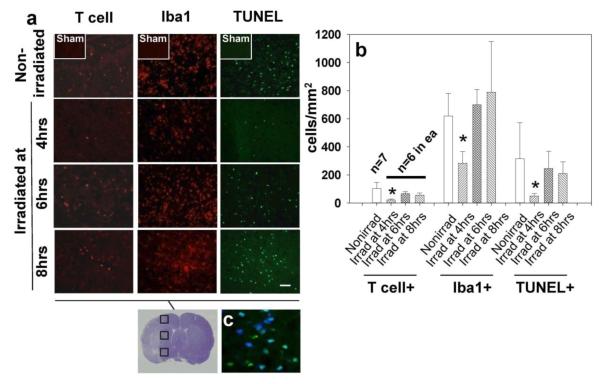
Fig. 4.
The effect of splenic irradiation on the immune invasion and inflammatory response in the post-ichemic brain. (a) Reduced number of invading T-cells, ameboid microglia, and apoptotic cells in the postischemic brain after splenic irradiation (magnification bar: 30μm). (b) Graph showing significantly reduced cell counts associated with irradiation at 4 hrs but not at 6 and 8 hrs after ischemia; *p<0.05 vs. nonirradiated, ANOVA. (c) The photograph (blown up X3) showing co-localization of TUNEL and neuronal marker, NeuN (blue color: NeuN, AMCA; green: TUNEL, fluorescein. Turquoise color results from merging AMCA and fluorescein component images)

