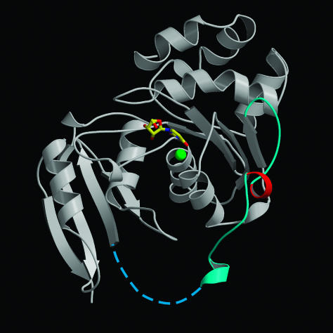
Fig. 5.
Ribbon representation of E. coli cytidine deaminase. Asp-128 maps to a helical turn (red) situated on a loop (cyan and dotted line) connecting the catalytic and pseudocatalytic domains of the E. coli enzyme. Uridine at the active site is shown in bonds representation. The green sphere represents Zn2+.

