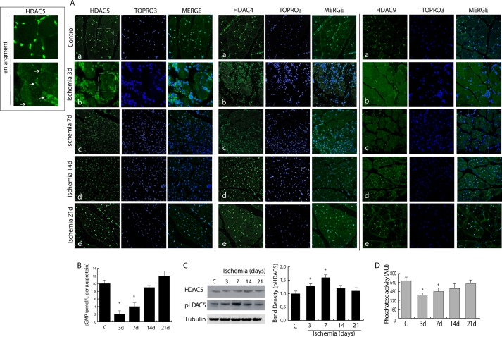
FIGURE 1.
Alteration in the dystrophin-nitric oxide pathway following ischemia determines class IIa nuclear export. A, confocal microscopy showing HDAC5, HDAC4, and HDAC9 distribution (green) in control (a) and post-ischemic adductor muscle at 3 (b), 7 (c), 14 (d), and 21 days (e). Nuclei were counterstained with TOPRO3 (blue). Magnification, ×40. Insets show HDAC5 distribution from control and ischemia 3-day panels; arrows indicate nuclei with low HDAC5 content. B, measurement of cGMP levels during ischemia. C, Western blotting showing the level of HDAC5 and its phosphorylation in control and in ischemic muscle. The graph shows the relative increase in HDAC5 phosphorylation normalized to total HDAC5 protein and to loading control (Tubulin). D, phosphatase activity in control and ischemic muscles. *, p < 0.05 versus control. Error bars, S.E. AU, arbitrary units.

