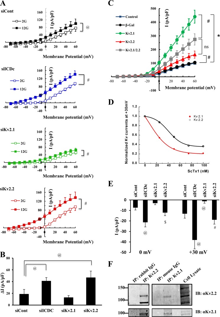
FIGURE 6.
Interactions of Kv2.2 and Kv2.1 regulate outward K+ current in β-cells. 832/13 cells were transfected with siControl (siCont), siICDc, siKv2.1, or siKv2.2 duplexes in combination with a plasmid encoding GFP and cultured for 72 h prior to patch clamping in a Warner chamber. A, normalized current-voltage relationships at 2 mm glucose (open symbols) and 12 mm glucose (closed symbols). B, average glucose-stimulated K+ current at 0 mV. Data shown are average ± S.E. (error bars) of 5–12 cells patched per condition. #, p < 0.01, @, p < 0.05. For C and D, 832/13 cells were either left untreated or treated with adenoviruses expressing β-gal, Kv2.1, Kv2.2, or combinations thereof and cultured for 24 h prior to patch clamp analyses. C, data shown are average ± S.E. (error bars) of 7–21 cells patched per condition. *, p < 0.001; #, p < 0.01 at a membrane potential of 0 mV. D, ScTx1 dose-response curve in 832/13 cells overexpressing Kv2.1 or Kv2.2 by adenovirus-mediated gene transfer. E, inhibitory effects of ScTx (25 nm) on outward K+ current at 0 and +30 mV in 832/13 cells treated with siICDc, siKv2.1, siKv2.2, or siControl. Data shown are average ± S.E. (error bars) of 5–7 cells patched per condition. @, p < 0.05 compared with siControl; $, p < 0.05 compared with siKv2.1; #, p < 0.001 compared with siKv2.1. F, 832/13 cell lysates (native cells; no viral treatment) were immunoprecipitated (IP) by either anti-Kv2.2, anti-Kv2.1, or control IgG antibody. Immunoblots (IB) were probed with antibody specific for Kv2.2 or Kv2.1. Results are representative of three independent experiments.

