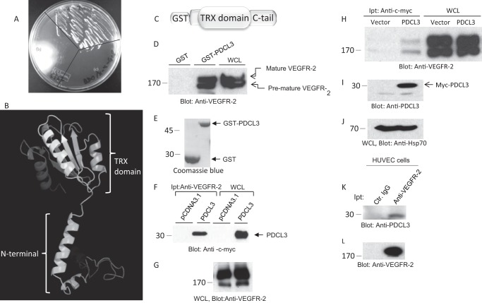
FIGURE 1.
Identification of PDCL3 as a VEGFR-2 binding protein. pGADT7-PDCL3 was transformed into Y187 yeast cells, and PGBKT7-VEGFR-2 was transformed into AH109 yeast cells. The two lines were mated and plated on QDO/X-α-Gal medium along with their haploid constituents. A, pGADT7-PDCL3/pGBKT7-VEGFR-2 diploids (a), pGBKT7-VEGFR-2 AH109 (b), and pGADT7-PDCL3/Y187 (c). B, the predicted structure of PDCL3. TRX, thioredoxin. C, schematic of the GST-PDCL3 fusion protein. D, cell lysates derived from PAE cells expressing VEGFR-2 were subjected to in vitro GST pull-down assay. WCL, whole cell lysate. E, Coomassie Blue stain of GST and GST-PDCL3. F, HEK-293 cells cotransfected with VEGFR-2 and an empty vector or PDCL3 were lysed, immunoprecipitated (Ipt) with anti-VEGFR-2 antibody, and blotted with anti-c-myc antibody. G, whole cell lysates from the same group were probed with anti-VEGFR-2 antibody. H, HEK-293 cells cotransfected with VEGFR-2 and an empty vector or PDCL3. Cells were lysed, immunoprecipitated with anti-c-myc antibody, and blotted with anti-VEGFR-2 antibody. Whole cell lysates from the same group were probed with anti-PDCL3 antibody (I) and anti-HSP70 antibody (J). K, cell lysates derived from HUVECs immunoprecipitated with control IgG or anti-VEGFR-2 antibody and blotted with an anti-PDCL3 antibody. L, the same membrane was reblotted with anti-VEGFR-2 antibody. All Western blot analyses and GST-pull down experiments were repeated at least three times.

