FIGURE 2.
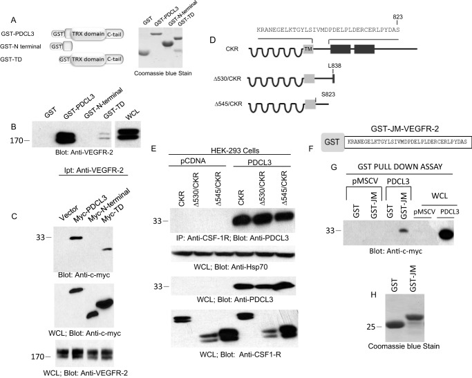
Juxtamembrane of VEGFR-2 is required for interaction with PDCL3. A, schematic of GST-PDCL3, GST-N terminus PDCL3, and GST-thioredoxin domain (TD) and the Coomassie Blue stain of recombinant proteins purified from E. coli. TRX, thioredoxin. B, cell lysates derived from HEK-293 expressing VEGFR-2 subjected to an in vitro GST-pull down assay using GST alone or GST-PDCL3, GST-N terminus PDCL3, and GST-TD. WCL, whole cell lysate. C, top panel, HEK-293 cells cotransfected with an empty vector, PDCL3, or truncated PDCL3 constructs with VEGFR-2 were lysed, immunoprecipitated (Ipt) with anti-VEGFR-2 antibody, and blotted with anti-c-myc antibody. Whole cell lysates derived from the same group were blotted with anti-c-myc antibody and anti-VEGFR-2 antibody. D, schematic of carboxyl domain-deleted chimeric VEGFR-2 (CKR). HEK-293 cells were engineered to express wild-type CKR and truncated CKR constructs alone or coexpress with PDCL3. E, top panel, cells were lysed and subjected to coimmunoprecipitation (IP) using anti-CSF-1R antibody that specifically recognizes the extracellular domain, followed by blotting with anti-PDCL3 antibody. Whole cell lysates were blotted for protein loading control, anti-Hsp70 antibody, anti-PDCL3 antibody, and anti-VEGFR-2 antibody. F, schematic of the GST-JM (juxtamembrane) domain of VEGFR-2. G, the GST-JM domain of VEGFR-2 was incubated with PAE cell lysates expressing GST or GST-PDCL3, subjected to GST-pull down assay, and blotted with anti-c-myc antibody. H, Coomassie Blue stain of GST and the GST-JM domain of VEGFR-2. The blots are representative of at least three independent experiments.

