FIGURE 3.
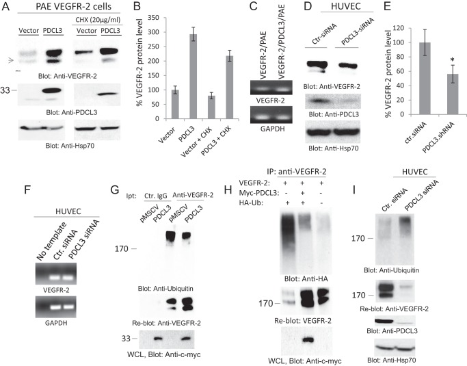
PDCL3 regulates the abundance of VEGFR-2 protein through inhibition of VEGFR-2 ubiquitination. A, PAE cells coexpressing VEGFR-2 with an empty vector or PDCL3 were treated with vehicle or cycloheximide (CHX) (20 ng/ml, 90 min). Cells were washed and lysed. Whole cell lysates were subjected to Western blot analysis using anti-VEGFR-2 antibody, anti-PDCL3 antibody, and anti-HSP70 antibody. B, graph representing three independent experiments. C, the mRNA derived from PAE cells expressing VEGFR-2 alone and coexpressing VEGFR-2 with PDCL3 were subjected to RT-PCR analysis using PCR primers for VEGFR-2 or GAPDH. D, HUVECs were transfected with either control siRNA (Ctr-siRNA) or PDCL3 siRNA. After 48 h, cells were lysed, and whole cell lysates were blotted for anti-VEGFR-2 antibody, anti-PDCL3 antibody, and anti-Hsp70 antibody. E, graph representing three independent experiments. *, p < 0.031. F, the mRNA derived from HUVECs transfected with the control siRNA and PDCL3 siRNA was subjected RT-PCR analysis using PCR primers for human VEGFR-2 or GAPDH. G, top panel, PAE cells coexpressing VEGFR-2 with an empty vector (pMSCV) or PDCL3 were washed, lysed, immunoprecipitated (Ipt) with anti-VEGFR-2 antibody, and blotted with anti-ubiquitin antibody. The same membrane was reblotted with anti-VEGFR-2 antibody (center panel). Whole cell lysate (WCL) was subjected to Western blot analysis using anti-c-myc antibody for PDCL3 (bottom panel). H, HEK-293 cells were transfected with VEGFR-2 alone, VEGFR-2 with PDCL3, or VEGFR-2 with PDCL3 and HA-tagged ubiquitin (HA-Ub) (top panel). Cells were lysed, immunoprecipitated (IP) with anti-VEGFR-2 antibody, and blotted with anti-HA antibody. The same membrane was blotted with anti-VEGFR-2 antibody (center panel). Whole cell lysate from the same group was blotted for PDCL3 using anti-c-myc antibody (bottom panel). I, HUVECs were transfected with control or PDCL3 siRNA. Total cell lysates were subjected to immunoprecipitation using anti-ubiquitin antibody (top panel). The same membrane was reblotted with anti-VEGFR-2 antibody (center panel). Whole cell lysates were subjected to Western blot analysis using anti-PDCL3 antibody and anti-HSP70 antibody. All the Western blot analyses shown are the representative of at least three independent experiments.

