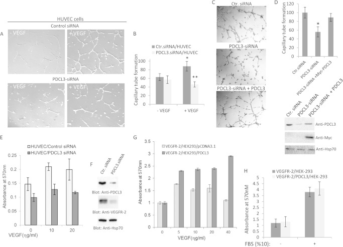
FIGURE 7.
PDCL3 activity is required for endothelial cell proliferation and angiogenesis. A, HUVECs were transfected with control (Ctr.) or PDCL3 siRNA, and after 16 h, cells were seeded onto Matrigel and stimulated with VEGF or vehicle. Cells were photographed after 16 h. B, the graph is the representative of four different fields and is presented as the mean ± S.D. *, p < 0.05; **, p < 0.021. C, HUVECs were transfected with control (Ctr.), PDCL3 siRNA alone or cotransfected with siRNA PDCL3 with c-Myc-PDCL3. Cells then were subjected to Matrigel tubulogenesis assay as in A. D, the graph is representative of four different fields and is presented as the mean ± S.D. *, p < 0.001. Cells lysates from the same group were blotted for PDCL3 and Hsp70 for protein loading control. E, HUVECs transfected with control siRNA or PDCL3 siRNA were subjected to a proliferation assay in the presence or absence of VEGF. F, whole cell lysates from the same siRNA-transfected HUVECs were subjected to Western blot analysis using anti-PDCL3 antibody and anti-Hsp70 antibody for protein levels. G, HEK-293 cells expressing VEGFR-2 were transfected with an empty vector or PDCL3. Cells were subjected to a proliferation assay and stimulated with varying amounts of VEGF. The proliferation of HEK-293 cells expressing VEGFR-2 alone or coexpressing VEGFR-2 with PDCL3 cells either kept in serum-free DMEM or stimulated with 10% FBS was measured using a 3-(4,5-dimethylthiazol-2-yl)-2,5-diphenyltetrazolium bromide assay as in E. H, the graph represents the quadruple of the mean ± S.D.

