FIGURE 2.

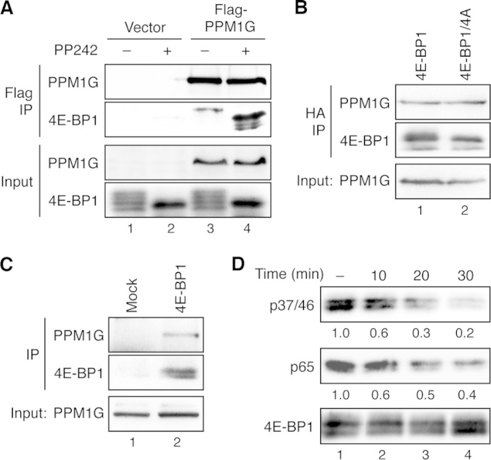
4E-BP1 is a direct substrate of PPM1G. A, PPM1G interacts with 4E-BP1 in cells. Stable HCT116 cells overexpressing HA-4E-BP1 were transfected with vector (lanes 1–2) or FLAG-PPM1G (lanes 3–4), and the transfected cells were treated with dimethyl sulfoxide or PP242 for 2 h. Cell lysates were prepared and immunoprecipitated (IP) with the anti-FLAG affinity gel. The presence of PPM1G in immunoprecipitates and cell lysates (10% input) was detected using the PPM1G antibody, whereas 4E-BP1 proteins were detected by the 4E-BP1 antibody. B, PPM1G interacts with both the WT and phosphorylation-deficient mutant 4E-BP1 in cells. Cell lysates were prepared from stable HCT116 cells overexpressing either WT HA-4E-BP1 or the HA-4E-BP1/4A mutant and subjected to immunoprecipitation using the anti-HA affinity matrix. The presence of PPM1G in immunoprecipitates and cell lysates (10% input) was detected using the PPM1G antibody, whereas 4E-BP1 proteins were detected by the 4E-BP1 antibody. C, the endogenous PPM1G and 4E-BP1 interact. Equal amounts of 293E cell lysates were incubated with protein A/G beads alone (Mock) or beads plus the 4E-BP1 antibody. The presence of PPM1G and 4E-BP1 in the immunoprecipitates and input were detected with the PPM1G and 4E-BP1 antibodies, respectively. D, dephosphorylation of 4E-BP1 in vitro. The HA-tagged 4E-BP1 was immunoprecipitated from stable HCT116/4E-BP1 cells using the anti-HA affinity matrix. Dephosphorylation reactions were carried out by incubating the immunoprecipitates with the purified PPM1G for the indicated time (lanes 2–4). As control for nonspecific dephosphorylation, HA-4E-BP1-bound beads were incubated in the dephosphorylation buffer without adding purified PPM1G for 30 min at room temperature (lane 1). Phosphorylation of 4E-BP1 at the Thr-37/46 and Ser-65 sites was detected using the phosphospecific antibodies specific for these sites. The relative phosphorylation of 4E-BP1 was quantified by normalizing ECL signals generated by phosphospecific antibodies to that of total 4E-BP1 and shown below the corresponding blots.
