
FIGURE 3.
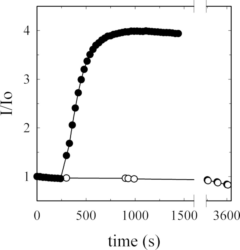
G-actin monomer stability during surface plasmon resonance analysis. G-actin tendency to aggregate or polymerize was assessed by measuring the fluorescence signal associated with pyrene-actin during incorporation into a filament (λex = 365 ± 3; λem = 407 ± 10, in nm). The fluorescence signal of 10 μm G-actin, containing 6% pyrene-label (○), was measured in a medium composed of 2 mm Tris-HCl (pH 7.7 at 25 °C), 70 μm CaCl2, and 0.005% C12E10. No changes in the intensity of fluorescence could be detected during 1 h. The slightly negative slope represents pyrene bleaching. As a positive control of actin polymerization, a 10× polymerization buffer was added to another aliquot of the preparation (●). The data are displayed as I/I0 (fluorescence signal at time t divided by initial fluorescence).