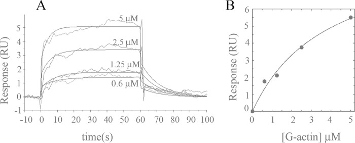
FIGURE 4.
Binding of G-actin to immobilized PMCA isolated from human erythrocytes. A, representative sensorgram of G-actin binding to immobilized PMCA at an immobilization level of ∼1000 RU is shown. PMCA was stabilized on the sensor surface in a micellar environment provided by the extraction detergent C12E10. Immediately after immobilization, a buffer containing 0.005% C12E10 was injected. G-actin was assayed in the range 0.6–5 μm in a modified Buffer G supplemented with 0.005% C12E10. The time for the association phase was set to 60 s. Curves were corrected for bulk effects by simple subtraction of the corresponding control sensorgrams. B, binding interaction analyzed from the kinetic global fit assuming a 1:1 interaction fit. The Kd values that best fit the experimental data were 3.8 ± 1.2 μm. Dark gray lines represent the experimental curves, and continuous black lines are the corresponding fits.

