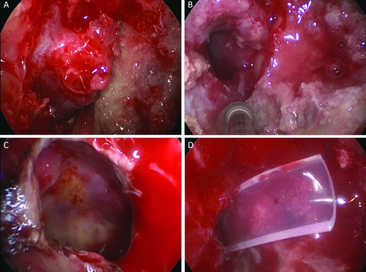
Figure 3.
Intraoperative view of the repeat transsphenoidal and infrapetrous approach. (A) Exposure of the petrous apex and cyst on a large surface. (B) Widening of the bony opening inferior to the petrous carotid into the petrous apex in a medial and inferior direction. (C) Visualization of the emptied cavity with a 45-degree endoscope. (D) Insertion of a Doyle splint into the cyst's cavity.

