
Fig. 2.
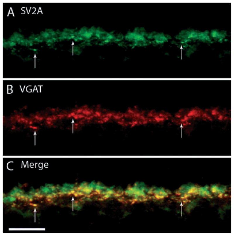
SV2A, a synaptic vesicle protein, labels horizontal cell terminals. A vertical section of guinea pig retina was double labeled with antibodies to SV2A and VGAT. (A) SV2A immunolabeling was localized to horizontal cell endings in the OPL, as well as photoreceptor terminals. (B) VGAT antibodies labeled horizontal cell terminals in the outer retina. (C) Merged images reveal the co-localization of SV2A labeling with that of VGAT. Arrows point to co-localized puncta along the OPL. Confocal images were scanned at 0.5 μm intervals and a total of eight optical images were obtained and compressed for viewing. Scale bar: 10 μm.