Fig. 3.
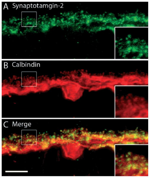
Synaptotagmin-2, a sensor for Ca2+-triggered vesicular release, localizes to horizontal cell processes and their terminals. A vertical section of guinea pig retina was double labeled with antibodies to synaptotagmin-2 and CaBP. (A) Synaptotagmin-2 antibodies labeled the processes in the OPL, with more intensely labeled dots throughout OPL. (B) CaBP immunostaining is in horizontal cell somata, processes and horizontal cell endings. (C) Merged images show co-localization of synaptotagmin-2 and CaBP immunostaining in horizontal cell processes and endings. The inset reveals a digital magnification of the boxed region indicating horizontal cell processes extending from the OPL. Confocal images were scanned at 0.7 μm intervals and a total of 10 optical images were obtained and compressed for viewing. Scale bar: 10 μm.

