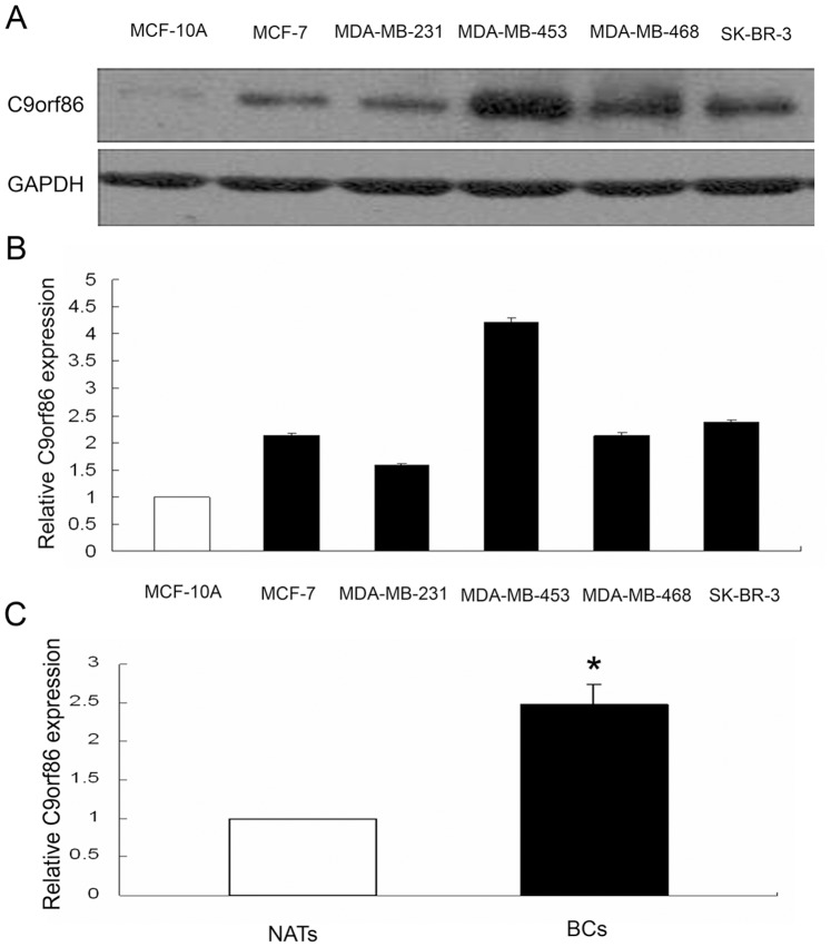
Figure 1. C9orf86 expression in breast cancer cells and tissues.
Expression of C9orf86 was quantified in human breast cancer (lanes 2–6), and normal (lane 1) breast epithelial cells by Western blot (A) and qRT-PCR (B). (C) QRT-PCR shows that expression of C9orf86 is increased in invasive BC tissues compared with NATs (P<0.05). Western blotting and RT-PCR were performed using glyceraldehyde-3-phosphate dehydrogenase (GAPDH) as a control.

