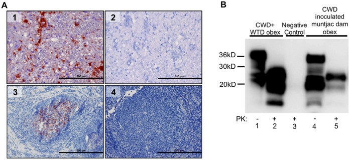
Figure 2. PrPCWD detection in CWD-inoculated dams.
(A) IHC PrPCWD is demonstrated (red deposits) in a tonsilar lymphoid biopsy collected at 4 mpi (A3) and in the obex of a terminal TSE diseased dam at 23 mpi (A1). Absence of red deposits is shown in negative control tonsil (A4) and obex (A2). Picture objective is 20X (scale bar = 200 µm). (B) Western blot detection of PrPCWD in obex tissue of a CWD-inoculated dam at 23 mpi (B lane 5) following PK digestion. Complete PK digestion of PrPC is shown in negative control deer obex (B lane 3). PrPCWD detection is demonstrated in CWD positive control deer obex (B lane 2).

