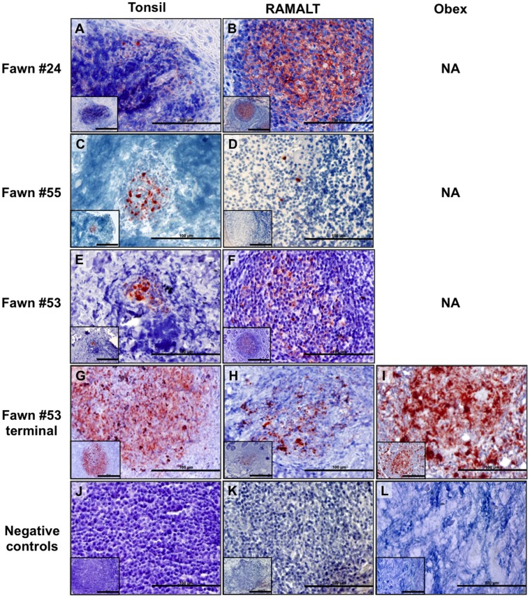
Figure 3. IHC PrPCWD detection in viable muntjac fawns born to CWD+ dams.
PrPCWD is demonstrated (red deposits) in tonsilar biopsies collected at 40 days post birth (dpb, A), 465 dpb (C) and 504 dpb (E) and RAMALT biopsies collected at 103 dpb (B), 386 dpb (D) and 504 dpb (F) and confirmed in the terminal tissues of 1 fawn (G, H, I). Absence of PrPCWD is shown in negative control tissues (J, K, L). Picture objective is 40X (scale bar = 100 µm) with insets at 20X (scale bar = 200 µm).

