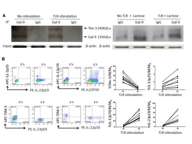
Figure 5. Activation of IL-12/IL-23 expressions by M/MØ following TLR stimulation and Tim-3/Gal-9 cis association.

A) Detection of Tim-3/Gal-9 physical association in un-stimulated and TLR-stimulated M/MØ by immunoprecipitation. The purified M/MØ were stimulated with or without LPS/R848 for 6 h; the procedure for co-immunoprecipitation of Tim-3 (40 Kd) and Gal-9 (35 Kd) was described in the Methods. Samples were pulled down by anti-Gal-9 or IgG antibodies and protein A/G PLUS-agarose; then probed with anti-Tim-3 and HRP-secondary antibody. β-actin was used to probe cell lysates for equal protein inputs. Repeated co-ip experiment by M/MØ in the presence of α-lactose in the cell lysates to inhibit random Gal-9 bound of Tim-3 are shown in the right panel. B) Activation of IL-12/IL-23 expressions by M/MØ following TLR stimulation. Purified M/MØ were cultured in the presence or absence of LPS/R848 for 6 h. Tim-3 cell surface expression and intracellular IL-12p35, IL-12p70, IL-23p19 productions were analyzed by flow cytometry as described in the Methods. Representative dot plots of the relationship between IL-12p35 and IL-23p19, IL-12p70 and IL-23p19, Tim-3 and IL-23p19, Tim-3 and IL-12p70, and summary data derived from multiple subjects were shown. Each line-linked symbol represents one subject’s M/MØ before and after TLR stimulation.
