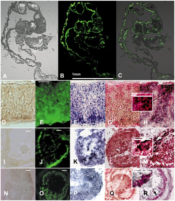
Figure 9. Brain stem cells appear to be multipotent in chick embryo chimeras.
Cells were first labeled genetically with a lentivirus expressing GFP under a CMV promoter. Cells were transplanted into the primitive streak of 20 h chick embryos. Top: Descendant cells in embryos harvested at two days later visible under blue excitation as green cells in a variety of tissue locations (C is an overlay of the image in A and the image in B) (HB: hind brain; MB: mid brain; E: eye; H: heart, LB: limb bud). Antibodies against phenotypic markers of the relevant tissues were used to confirm appropriate differentiation of heart muscle (CTNI), striated muscle (sα-actin), and neuronal cells (NF200) (pink stain; G, L, Q). Regions that had shown GFP-positive green human-descended cells (E, J, O) were then also processed for anti GFP-immunochemistry (black stain, F, K, P). As well some sections from regions processed for phenotypic markers (pink) were subsequently co-stained for GFP antigen (black, H, M, R). Insets of high magnification of 100 micron squares depict close proximity of black and pink staining. The black anti-GFP is perinuclear in pattern thus making it highly likely that the two markers are expressed together in individual cells. Panels D, I and N show no primary antibody controls for immunochemistry. Magnification: bars in B, 1 mm; D to R, 100 µm.

