
Figure 5. Dorsal flexion as a consequence of laterally flexing the posterior cervical vertebrae of Struthio camelus.
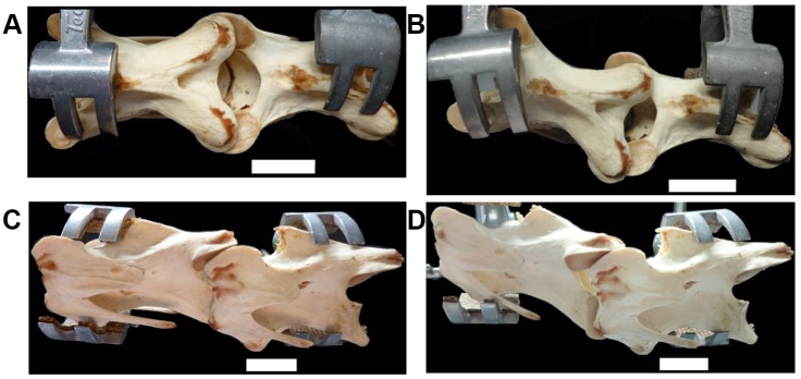
(a, c) C15 and C16 with no lateral flexion, and flexed ventrally to reach a dorsoventral angle of 0° (see zygapophyseal overlap (a)). (b, d) C15 and C16 flexed laterally, forcing dorsal flexion. Scale bars = 2cm.