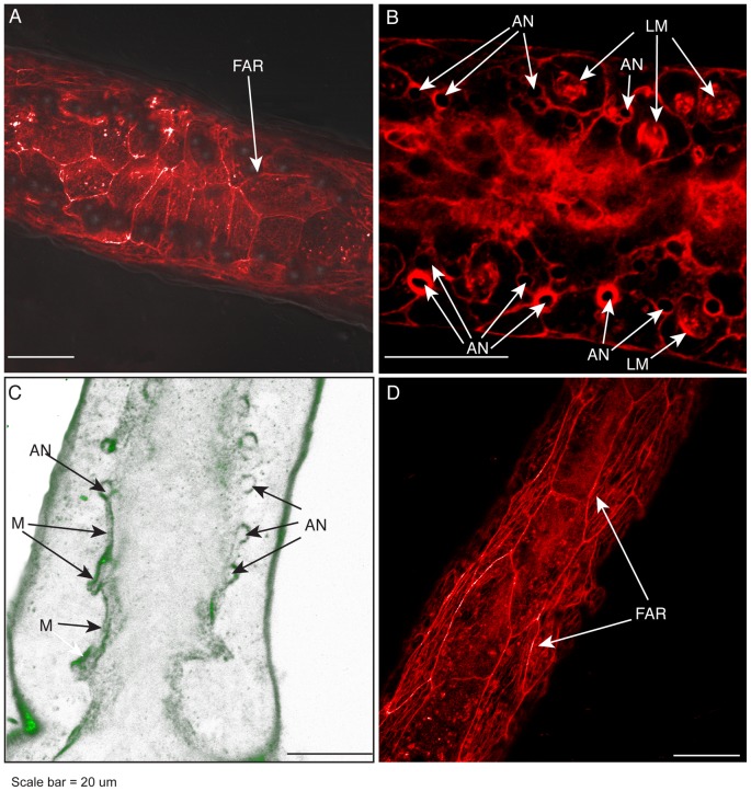
Figure 4. Basal epidermal actin, anchors, and mesoglea.
(A) Phalloidin stained F-actin rings in the basal epidermis beneath a polyp-stolon junction. (B) Phalloidin stained F-actin fibers surrounding some, but not all, anchors. Diffuse staining in the center of stolon is gastrodermal. (C) Immunohistochemical staining of collagen IV showing mesoglea (M) overlying and surrounding anchors. Optical depth, number of sections: 3 µm, 4. (D) Phalloidin stained F-actin rings in basal epidermis of stolon at positions proximal to the stolon top. Scale: 20 µm. AN, anchors; FAR, F-actin ring; LM, longitudinal muscle strands; M, mesoglea.

