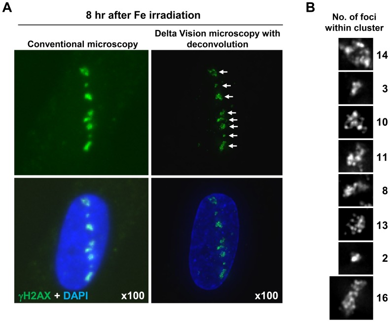
Figure 2. High resolution microscope analysis revealed clustered γH2AX foci formation within the tracks following Fe irradiation.
(A, B) Clustered γH2AX foci formation in 48BR (WT) primary G0/G1 cells is visualised using deconvolution. Images were captured with an Applied Precision DeltaVision RT Olympus IX70 microscope with deconvolution (right). The image resolution using the DeltaVision is compared with Zeiss Axioplan microscope without deonvolution (left). Enlarged individual clustered foci are shown with grey scale in B.

