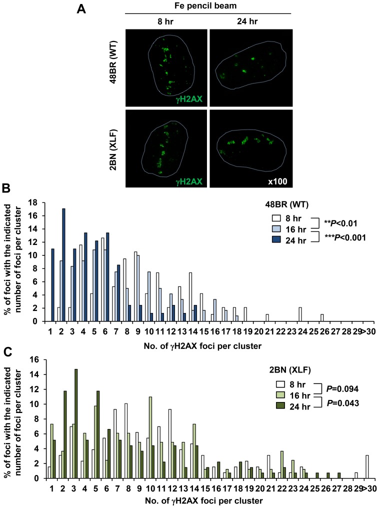
Figure 3. Clustered γH2AX foci arising within the particle tracks represent a signature of high LET particle radiation and are repaired slowly by NHEJ in G1.
(A) 48BR (WT) primary and 2BN (XLF) hTERT G0/G1 cells were irradiated in a horizontal direction with 1 Gy Fe irradiation. Images are taken using the DeltaVision microscope followed by deconvolution. Representative images at 8 and 24 h post Fe irradiation are shown. The nucleus outline is drawn with a dashed line from the DAPI staining. (B, C) Percentage of individual foci within a cluster was analysed from >100 individual clusters at each time point. Similar results were obtained in two independent experiments. Cells forming a single γH2AX track of length >8 µm and width >1 µm were analysed.

