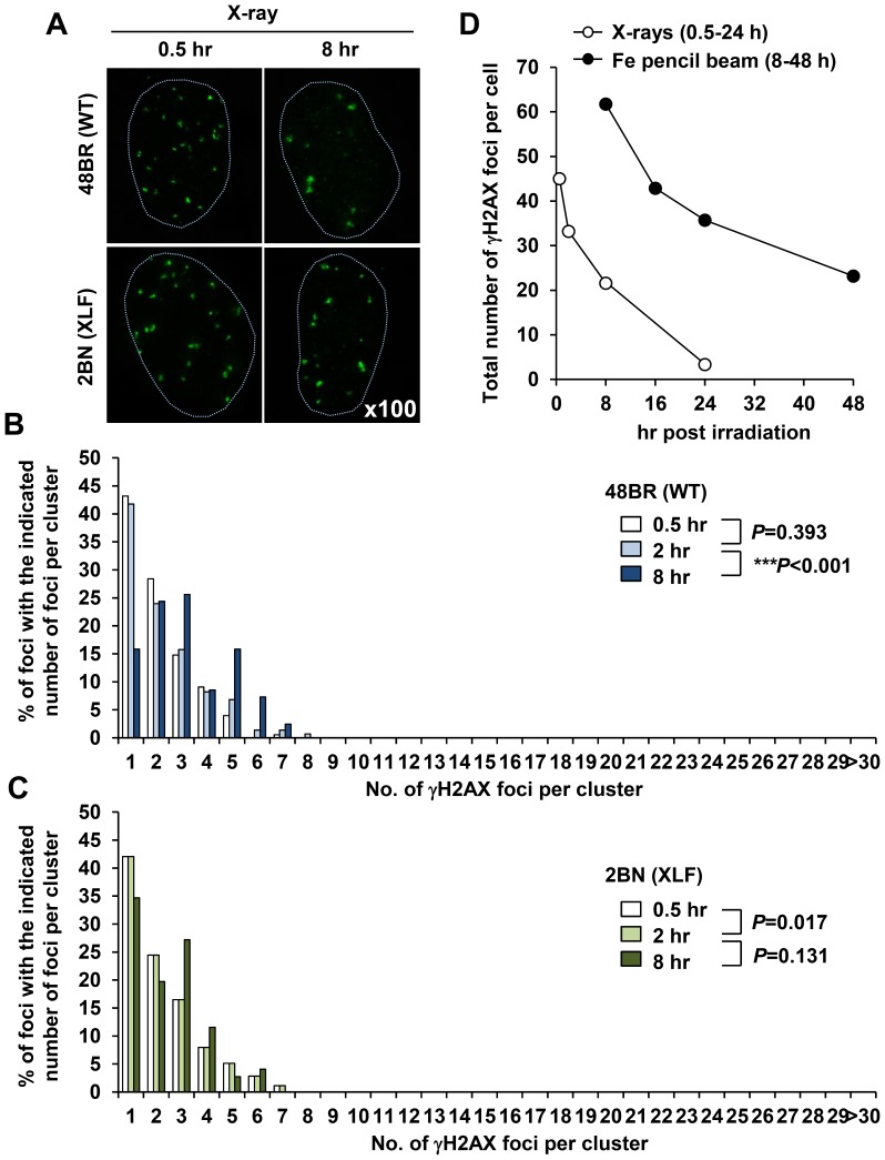
Figure 4. X-irradiation induces ‘Simple’ γH2AX foci.
(A) 48BR (WT) primary and 2BN (XLF) hTERT G0/G1 cells were irradiated with 1 Gy X-rays and stained with γH2AX and DAPI. Images are taken using the DeltaVision microscope followed by deconvolution. (B, C) Distribution of foci numbers in clusters was analysed from >100 individual clusters at each time point. (D) Repair kinetics following 1 Gy Fe ions is slower than that after 1 Gy X-rays. To examine repair kinetics, the total number of γH2AX foci within all the clusters per cell were enumerated from the cluster analysis in Figure 3B and 4B. Similar results were obtained in two independent experiments.

