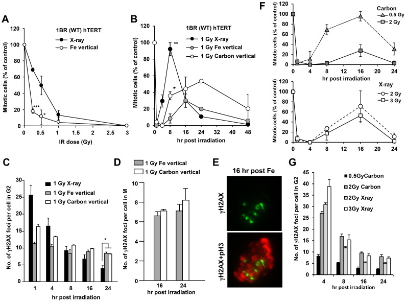
Figure 8. Cells with clustered γH2AX foci can enter mitosis despite prolonged checkpoint arrest following exposure to Fe ion irradiation.
A) Mitotic entry was examined in 1BR (WT) hTERT cells at 1 h post X-rays or vertical Fe irradiation. Cells, >400, were scored for mitotic index (MI). Mitotic cells were identified by p-histone H3 Ser10 and cellular morphology by DAPI. Results represent the MI relative to untreated cells. 1BR hTERT cells shows similar G2/M checkpoint responses to that in 48BR primary cells [40]. The size and number of γH2AX foci clusters between 48BR primary and 1BR hTERT cells were similar (data not shown). (B) The time of mitotic entry was examined following exposure to 1 Gy X-rays, Fe or Carbon ion irradiation. (C) The number of clustered γH2AX foci in G2 cells was enumerated by 2D analysis using normal microscopy following 1 Gy X-rays, Fe or Carbon ion irradiation. G2 cells were identified by CENP-F staining. (D) Since irradiated G2 cells restart cell cycle progression from 16 h post heavy ion irradiation, the number of DSBs in mitotic cells at 16 and 24 post IR was examined by scoring γH2AX foci in p-histone H3 Ser10 positive M phase cells. (E) Typical image of γH2AX foci in metaphase 1BR hTERT cells were taken by DeltaVision 3D imaging. (F) The maintenance of checkpoint arrest was examined following 0.5, 2 Gy Carbon or 2, 3 Gy X-rays. (G) γH2AX foci in G2 cells were enumerated following 2 Gy Carbon or X-rays. Mitotic index and γH2AX foci in G2 or M cells were examined by 2D analysis using Olympus BX51 or Zeiss Axioplan microscope.

