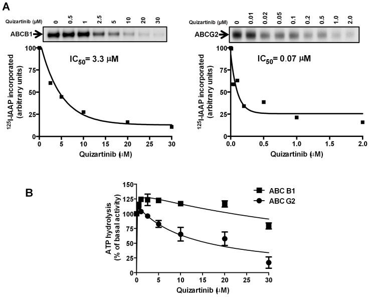
Figure 2. Quizartinib decreased [125I]-IAAP photolabeling of both ABCB1 and ABCG2 but inhibited ATPase activity of only ABCG2 at high concentrations.
Crude membranes from High-Five insect cells expressing ABCB1 and MCF-7 FLV1000 cells expressing ABCG2 were incubated with 0–30 µM quizartinib for 5 minutes at 21–23°C in 50 mM Tris-HCl, pH 7.5 and 3–6 nM [125I]-IAAP (2200 Ci/mmole) was added, followed by processing as described in Materials and Methods. (A) Representative autoradiograms from one experiment are shown in the upper panels; similar results were obtained in two additional experiments. In the lower panels, incorporation of [125I]-IAAP (from autoradiogram, Y-axis) into the ABCB1 and ABCG2 bands was plotted as a function of quizartinib concentration (X-axis). Quizartinib inhibited [125I]-IAAP binding to ABCB1 and ABCG2 with IC50’s of 3.3 µM and 0.07 µM, respectively, and the latter correspond to a therapeutically relevant plasma concentration. Values are from a representative experiment among three independent experiments. (B) Crude membrane protein from High Five insect cells expressing ABCB1 or ABCG2 was incubated with quizartinib at a range of concentrations in the presence or absence of sodium orthovanadate or BeFx (beryllium sulfate and sodium fluoride), respectively, as described in Materials and Methods. The ATPase activity in the presence of the indicated concentrations of quizartinib was calculated as a percent of basal (no addition of drug). The mean and standard error values from three independent experiments for ABCB1 (filled squares) and ABCG2 (filled circles) are shown.

