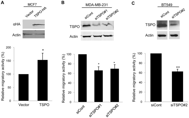
Figure 5. TSPO promotes breast cancer cell migration.
A. Control or TSPO expression vectors were transiently expressed in MCF7 cells. HA-tagged TSPO was detected by immunoblotting using an antibody against the HA tag, with actin as a loading control (top panel). The control and HA-TSPO-expressing MCF7 cells were allowed to migrate toward 10% FBS for 24 h in a transwell assay as described under Materials and Methods. Duplicate wells were used for each of three independent experiments. The results are expressed relative to the migration of control cells ( = 100%). Column (bottom panel): Mean of three experiments. Error bar: SD. P value was determined by Student's t-test. * p<0.05.indicates a significant difference between control and TSPO-overexpressing MCF7 cells. Control siRNA and siRNA sequences against TSPO (siTSPO #1 or siTSPO #2) were used to transfect MDA-MB-231 cells (B) or BT549 (C). The extent of silencing was determined by immunoblotting with antibodies against TSPO, with actin used as a loading control (top panel). Control and TSPO-depleted cells were then subjected to transwell assays as described under Materials and Methods Duplicate wells were used for each of three independent experiments. The results are expressed relative to the migration of control cells ( = 100%). Column (bottom panel): Mean of three experiments. Error bar: SD. P-value was determined by Student's t-test. * p<0.05 and ** p<0.01 indicate significant differences between control and TSPO-depleted cells.

