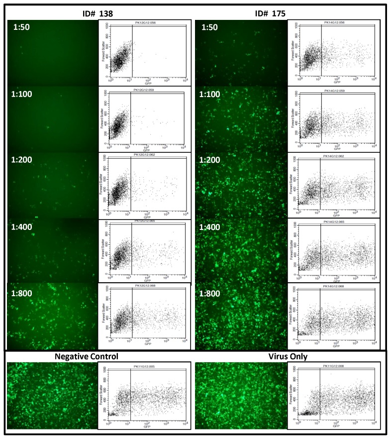
Figure 1. KSHV Neutralization Assay.
Fluorescent pictographs of 293 cells infected with rKSHV.219 incubated with serially diluted plasma samples (1∶50, 1∶100, 1∶200 and 1∶800) from two Kaposi’s sarcoma patients (ID 138 and 175) who were neutralizing antibody positive. ID 138 had comparatively higher titer neutralizing antibodies as compared to ID 175. Parallel flow cytometry scatter plots are also presented. Negative control (KSHV and KS negative plasma at a dilution of 1∶50) and virus only control are shown at extreme right.

