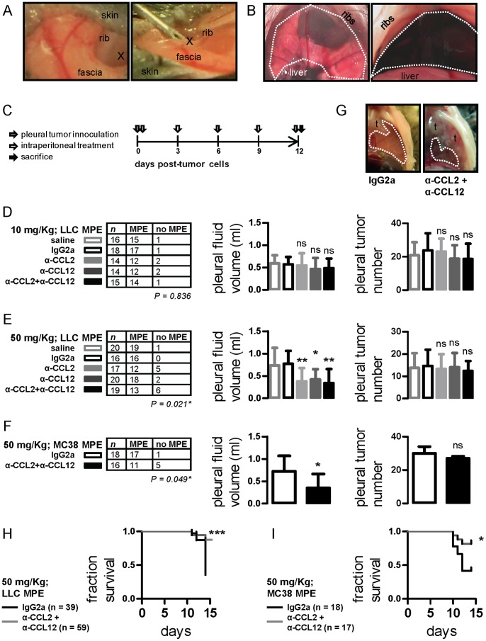
Figure 1. Impact of anti-CCL2 and/or anti-CCL12 monoclonal antibody treatment on syngeneic models of malignant pleural effusion (MPE).
(A) Photographs of intrapleural injection technique (x indicates the injection site). (B) Transdiaphragmatic photograph of mouse pleural space before (left) and four hours after (right) intrapleural Evans’ blue delivery. The dashed lines indicate the pleural confines. (C) Graphical outline of in vivo experiments. C57BL/6 mice received intrapleural Lewis lung carcinoma (LLC) or MC38 colon adenocarcinoma cells (grey arrow) followed by intraperitoneal treatment with normal saline, IgG2a, anti-CCL2, anti-CCL12, or anti-CCL2 plus anti-CCL12 every three days (white arrows). Mice were terminated after 12 days (black arrow). Primary end-points were MPE incidence and volume. Secondary end-points were pleural tumor number and survival. (D) MPE incidence (left), volume (middle), and pleural tumor number (right) of mice with LLC-induced MPE after regular-dosed antibody treatment. (E) MPE incidence (left), volume (middle), and pleural tumor number (right) of mice with LLC-induced MPE after high-dose antibody treatment. (F) MPE incidence (left), volume (middle), and pleural tumor number (right) of mice with MC38-induced MPE treated with high-dose IgG2a control or anti-CCL2/12 combination regimen. (G) Transdiaphragmatic photographs of representative MC38-induced MPEs from an IgG2a and an anti-CCL2/12 combination-treated mouse. Dashed lines outline MPEs and t designates pleural tumors. (H,I) Fractional survival of mice with LLC- and MC38-induced MPE. Columns, mean; bars, SD; n, sample size; P, probability by χ2 test; ns, *, **, ***: P>0.05, P<0.05, P<0.01, and P<0.001 compared with saline and/or IgG2a control.

