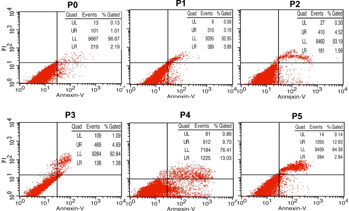
Figure 2. Apoptosis tests of primary KCs and subcultured cells.
The presence of apoptotic cells was identified by flow cytometric analysis of cells labeled with Annexin V and propidium iodide. Cells in the lower right quadrant correspond to early apoptotic cells (Annexin V-positive and propidium iodide-negative), while cells in the upper quadrant correspond to late apoptotic or necrotic cells (Annexin V-positive and propidium iodide-positive). The results showed that the percentage of apoptotic cells was increased over the course of passaging (especially after passage 3).

