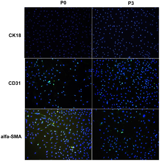
Figure 4. Immunofluorescence tests for contaminating cells (100×).
The results indicated that contaminating cells detected both in P0 and P3 cell cultures were few and included SECs (positive for CD31) and HSC (positive for alpha-SMA), but not hepatocytes (positive for CK18). (Blue: DAPI, Green: FITC beads).

