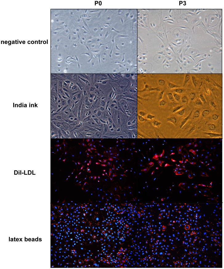
Figure 5. Phagocytic activity of kupffer cells (100×).
Both P0 and P3 cells displayed strong phagocytic activity: nearly all of the cells incorporated the ink, Dil-LDL and latex beads 4 h after administration. In addition, there were no significant differences in the phagocytic abilities of P0 and P3.

