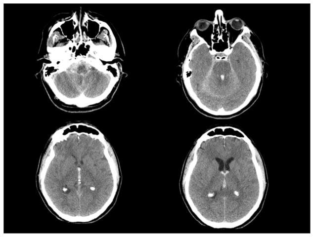
Fig. 1.
Axial CT scan of the head on presentation showing diffuse subarachnoid hemorrhage concentrated in the posterior fossa and foramen magnum. Intraventricular hemorrhage is most prominent in the fourth ventricle, third ventricle, extending into the bilateral lateral ventricles through the foramens of Monro.

