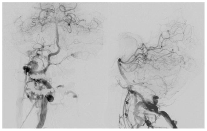
Fig. 2.
Initial angiogram (right vertebral injection) showing rapid arteriovenous shunting from the distal V-2 segment of the right vertebral artery through a single hole fistula draining into the high cervical venous structures. Late arterial phase images (left: anterioposterior projection, right: lateral projection) showing early and prominent opacification of the right suboccipital, paravertebral, clival (including contralateral), and epidural veins, as well as the marginal and condylar sinuses.

