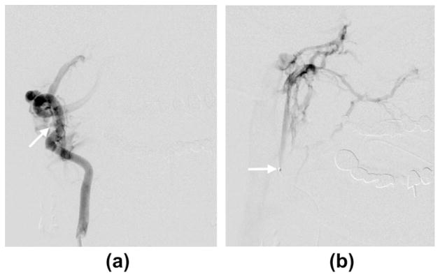
Fig. 3.

(a) Anteroposterior oblique view of right vertebral artery angiogram showing the fistulous connection (arrow). (b) The microcatheter traverses the fistula with its tip (arrow) in the venous compartment. Microcatheter injection showing ascending venous drainage into the skull base veins (magnified anterioposterior projection).
