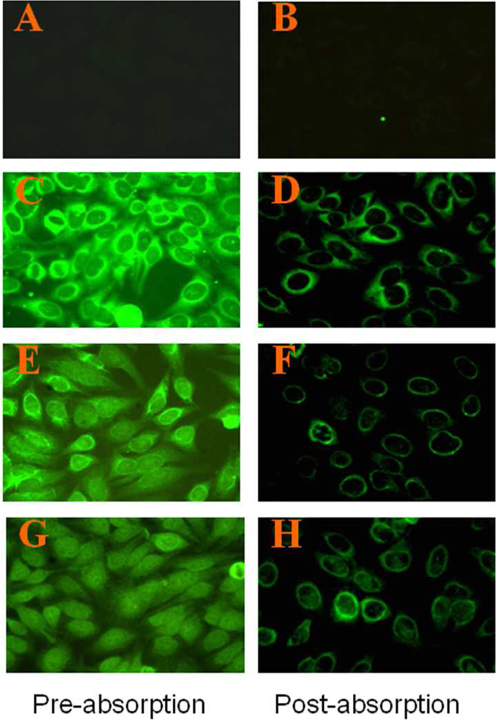
Figure 3. The 47-kDa protein is a cytoplasmic or perinuclear protein, as revealed by immunofluorescence analysis of HEp-2 cell substrate using human liver fibrosis sera.
Immunofluorescence was done using patients’ sera or sera purified with 47-kDa antigen (see Materials and Methods). Left panel (A, C, E, G): pre-absorption sera; right panel (B, D, F, H): post-absorption sera. Three representative liver fibrosis sera (D, F and H) were used, and one normal human serum (A) was used as control.

